Blocked arteries are one of the leading causes of serious cardiovascular problems, including heart attack and stroke. Detecting them early can significantly reduce health risks and allow for timely treatment. One of the most effective, non-invasive ways to identify arterial blockages is through a vascular ultrasound. This diagnostic imaging test provides a safe, painless, and highly accurate method of evaluating blood flow in your arteries and veins.
In this article, we’ll explore how vascular ultrasound works, why it is used to detect blocked arteries, the benefits of early detection, and what you can expect during the procedure.
What Is a Vascular Ultrasound?
A vascular ultrasound is a diagnostic imaging test that uses high-frequency sound waves to create real-time images of blood vessels. Unlike X-rays or CT scans, it does not use radiation. Instead, it relies on sound waves to evaluate the structure and function of veins and arteries.
There are two main types of vascular ultrasound:
Traditional Ultrasound – Provides images of the structure of blood vessels.
Doppler Ultrasound – Measures the movement of blood through the arteries and veins, showing the speed and direction of blood flow.
By combining both, vascular ultrasound helps physicians detect narrowing (stenosis), blockages, or blood clots that may affect circulation.
How Vascular Ultrasound Detects Blocked Arteries
Blocked arteries occur when fatty deposits (plaque) build up inside the vessel walls, reducing or completely stopping blood flow. This condition is known as atherosclerosis. Left untreated, it can cause life-threatening events such as a heart attack or stroke.
A vascular ultrasound helps detect blocked arteries in several ways:
Evaluating Blood Flow
Doppler technology shows how blood is moving through the arteries. Reduced or turbulent flow patterns often indicate narrowing or blockage.Measuring Arterial Narrowing
The ultrasound can visualize plaque buildup and measure the degree of stenosis. Even a partial blockage can be identified before it becomes dangerous.Detecting Blood Clots (Thrombosis)
Clots within arteries or veins can be detected early with ultrasound imaging, preventing complications like deep vein thrombosis (DVT) or pulmonary embolism.Monitoring Circulation in High-Risk Areas
Vascular ultrasound is often used to check arteries in the neck (carotid arteries), legs (peripheral arteries), and abdomen (aorta) for blockages that could cause serious health problems.
Common Conditions Diagnosed with Vascular Ultrasound
Vascular ultrasound is an essential tool in detecting and monitoring various cardiovascular conditions, including:
Carotid Artery Disease – Narrowing in the neck arteries that can lead to stroke.
Peripheral Artery Disease (PAD) – Blockages in the leg arteries that cause pain and reduced circulation.
Abdominal Aortic Aneurysm (AAA) – An enlarged area in the aorta that may rupture if not treated.
Deep Vein Thrombosis (DVT) – Blood clots in deep veins, usually in the legs.
Venous Insufficiency – Poor circulation due to damaged vein valves.
By identifying these conditions early, patients can receive treatment before complications arise.
Benefits of Detecting Blocked Arteries Early
Early detection of blocked arteries with a vascular ultrasound offers several benefits:
Prevention of Serious Complications
Identifying narrowing or blockages early helps prevent heart attack, stroke, or limb damage.Guiding Treatment Plans
Results from a vascular ultrasound allow doctors to decide whether lifestyle changes, medications, or procedures like angioplasty are necessary.Non-Invasive and Painless
Unlike surgical procedures or angiography, vascular ultrasound is safe, requires no incisions, and has no recovery time.Radiation-Free Imaging
Since it doesn’t use X-rays, it’s safe for repeated monitoring, even in pregnant women or patients requiring frequent follow-ups.Cost-Effective and Widely Available
Vascular ultrasound is more affordable than advanced imaging tests like MRI or CT scans, making it accessible for routine screenings.
Who Should Consider a Vascular Ultrasound?
Not everyone needs regular vascular ultrasounds, but certain groups are at higher risk for arterial blockages. You may benefit from this test if you have:
High blood pressure or high cholesterol
Diabetes
Smoking history
Obesity
A family history of heart disease or stroke
Leg pain, swelling, or cramping when walking
History of stroke or mini-stroke (TIA)
If you fall into one or more of these categories, your doctor may recommend a vascular ultrasound as part of your preventive heart health screening.
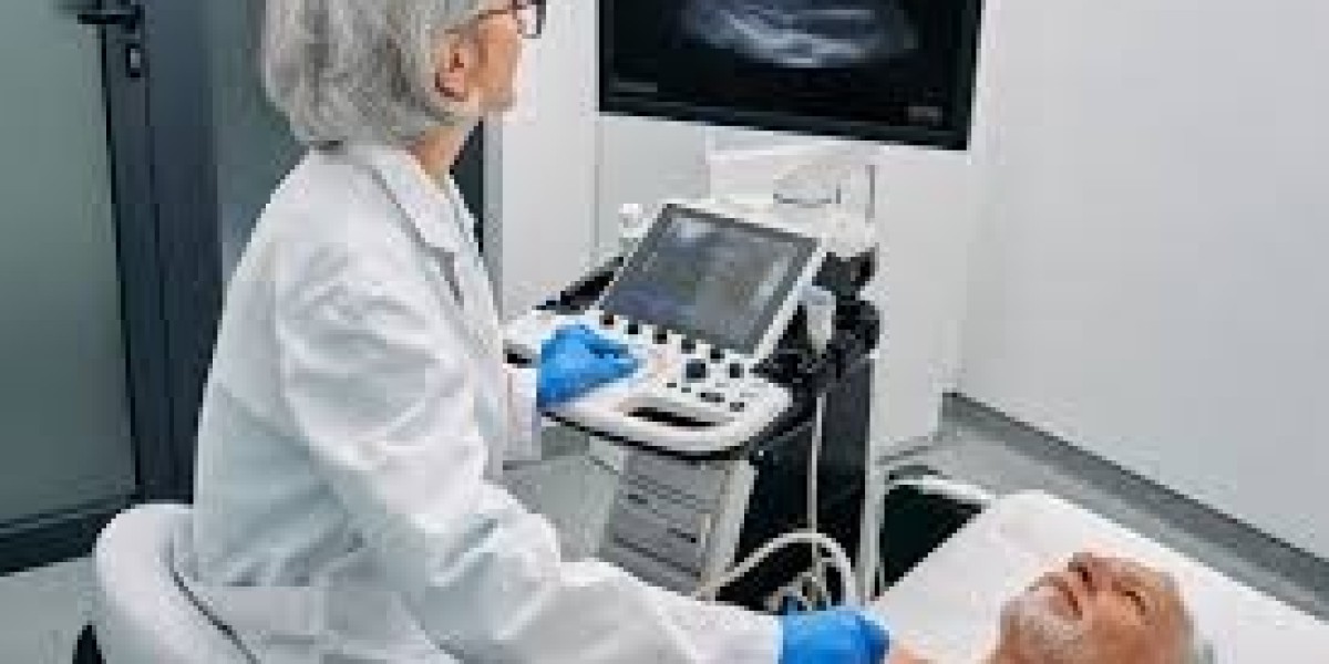
What to Expect During a Vascular Ultrasound
Many patients feel more comfortable knowing what the test involves. Here’s what typically happens:
Preparation
No special preparation is required for most vascular ultrasounds, although fasting may be necessary for abdominal scans.Procedure
You’ll lie on an exam table.
A technician applies a gel to the skin to help transmit sound waves.
A handheld device called a transducer is moved over the target area.
Real-time images of your blood vessels appear on a monitor.
Duration
The test usually takes 30–60 minutes.After the Test
You can immediately resume normal activities, and results are typically available within a short time.
Treatment Options After a Blockage Is Found
If a vascular ultrasound shows narrowing or blocked arteries, treatment depends on the severity:
Lifestyle Changes – Quitting smoking, eating a heart-healthy diet, and exercising regularly.
Medications – Blood thinners, cholesterol-lowering drugs, or blood pressure medications.
Minimally Invasive Procedures – Angioplasty, stenting, or atherectomy to open blocked arteries.
Surgery – In severe cases, bypass surgery may be recommended.
The goal is to restore proper blood flow and reduce the risk of life-threatening complications.
Final Thoughts
A vascular ultrasound is one of the most effective and accessible ways to detect blocked arteries early. It’s safe, painless, non-invasive, and provides doctors with crucial information about blood flow and circulation. Detecting arterial blockages before symptoms worsen can help prevent heart attacks, strokes, and other serious cardiovascular conditions.
If you are at risk for vascular disease or experiencing symptoms like leg pain, numbness, or dizziness, talk to your doctor about scheduling a vascular ultrasound. Early detection could save your life.